Table of Contents
- Introduction
- Traditional Impression Techniques
- Digital Impression Technology
- Accuracy Comparison
- Cost Analysis
- Workflow Efficiency
- Patient Experience
- Choosing the Right Method
- Conclusion
Introduction
The dental impression is the foundational step in indirect restorations—crowns, bridges, implants, and orthodontic appliances all depend on an accurate reproduction of the patient’s oral anatomy. For over a century, elastomeric impression materials (polyvinyl siloxane, polyether) served as the standard. The advent of digital impression technology through intraoral scanning has fundamentally transformed this workflow, prompting many practices to evaluate whether to transition from analog to digital.
This article provides an objective comparison of digital and traditional impression methods, examining accuracy, cost, workflow, and patient experience to help practices make informed decisions.
Traditional Impression Techniques
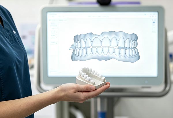
Traditional impressions involve loading a tray with elastomeric material, seating it in the patient’s mouth, and waiting for the material to polymerize. The resulting physical model is then poured in dental stone or shipped to a laboratory for fabrication.
Common Materials
- Polyvinyl siloxane (PVS): Excellent detail reproduction, dimensional stability, and hydrophilicity
- Polyether: Superior accuracy for implant impressions; hydrophilic; stiffer set
- Additional silicone: Cost-effective option for less critical applications
Strengths
- Well-established technique with decades of clinical validation
- Low equipment cost—no capital investment required
- Familiar to all dental professionals
- Capable of capturing full-arch impressions reliably
Weaknesses
- Patient discomfort—gag reflex, taste, claustrophobia
- Risk of distortion from tray movement, material voids, or temperature effects
- Cannot verify quality until the model is poured
- Requires physical storage and shipping
- Environmental sensitivity—humidity and temperature affect setting
Digital Impression Technology
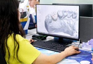
Digital impressions use intraoral scanners (IOS) to capture the oral anatomy as a three-dimensional point cloud, which is then converted into a highly accurate digital model (STL file). Leading systems include the iTero Element, 3Shape TRIOS, CEREC Primescan, and Medit i series.
How It Works
The scanner projects a light pattern (confocal microscopy, triangulation, or structured light) onto the tooth surface. Cameras capture the reflected pattern, and software algorithms calculate the 3D coordinates of millions of surface points. As the clinician moves the scanner wand across the arch, the software stitches consecutive captures into a seamless digital model in real time.
Strengths
- Immediate visual feedback—errors can be corrected on the spot
- Superior patient comfort—no impression material or trays
- Digital files transmit instantly to the laboratory
- Eliminates distortion risk from material shrinkage
- Integrates with CAD/CAM for same-visit restorations
- Stores indefinitely without physical space requirements
Weaknesses
- High initial equipment investment ($15,000–$50,000+)
- Learning curve for efficient scanning technique
- May struggle with blood, saliva, or subgingival margins
- Full-arch scanning can be time-consuming for edentulous cases
- Requires compatible laboratory infrastructure
Accuracy Comparison
Accuracy in dental impressions encompasses two dimensions: trueness (closeness to the actual anatomy) and precision (reproducibility of measurements). Multiple in vitro and clinical studies have compared digital and traditional methods:
- Single-unit crowns: Digital scans achieve comparable or superior accuracy to PVS impressions, with trueness values of 15–30 micrometers versus 20–40 micrometers for traditional methods.
- Short-span bridges (3–4 units): Digital scans maintain acceptable accuracy, though results depend heavily on scanner model and operator skill.
- Full-arch scans: Traditional impressions may still hold a slight edge in full-arch precision, though newer generation scanners are narrowing this gap significantly.
- Implant impressions: Digital scan bodies provide reliable results for single and short-span implant cases; full-arch implant digital workflows require careful technique and specific scan body systems.
The overall consensus in the current literature is that for most single-unit and short-span restorations, digital impressions are at least as accurate as traditional methods, with the added benefit of immediate quality verification.
Cost Analysis
| Cost Factor | Traditional | Digital |
|---|---|---|
| Initial investment | $200–$500 (trays, material stock) | $15,000–$50,000+ (scanner + software) |
| Per-impression material cost | $8–$25 (PVS/polyether) | $0–$5 (disposable scanner tips) |
| Model cost | $5–$15 (stone, die stone) | $2–$10 (3D printed model, optional) |
| Shipping | $5–$20 per case | $0 (digital transfer) |
| Remake cost (impression error) | High—patient recall required | Low—rescan immediately |
While the upfront cost of digital is substantial, practices with high case volumes often reach break-even within 12–24 months through material savings, reduced remakes, and improved workflow efficiency.
Workflow Efficiency
Digital impressions compress the restorative workflow significantly. Traditional workflows involve impression-taking, model pouring, shipping, lab fabrication, and a second patient visit. Digital workflows can eliminate model pouring and shipping, enable electronic lab prescriptions, and even support same-day CAD/CAM fabrication.
Key efficiency gains include:
- Reduced chair time (5–8 minutes for a digital scan vs. 10–15 minutes for traditional)
- Elimination of model pouring and physical shipping
- Faster lab turnaround (files received same day)
- Fewer remakes due to impression errors
- Better lab communication through digital annotations
Patient Experience
Patients consistently report preferring digital impressions over traditional methods. Key reasons include:
- No unpleasant-tasting impression material
- No gag reflex trigger from bulky trays
- Ability to see their teeth on screen in real time
- Shorter appointment duration
- Perception of modern, technology-driven care
For practices focused on patient experience marketing, digital scanning can be a significant differentiator and referral driver.
Choosing the Right Method
Most practices benefit from a hybrid approach during transition:
- Use digital for: Single crowns, short bridges, inlays/onlays, orthodontic scans, and patient communication
- Use traditional for: Full-arch implant cases (until digital workflows mature), cases with deep subgingival margins, and when scanner is unavailable
Conclusion
Digital impression technology represents a genuine paradigm shift in dental practice. While the initial investment requires careful financial analysis, the combined benefits of accuracy, patient comfort, workflow efficiency, and reduced remakes make digital scanning an increasingly compelling choice. Practices that adopt digital workflows position themselves for the future of dentistry while delivering a superior patient experience today.
The traditional impression technique remains a reliable fallback and still holds advantages in specific clinical scenarios. However, as scanner technology continues to improve and costs decrease, the transition to fully digital impression workflows appears inevitable for most modern dental practices.